Fig. 3.
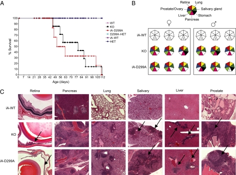
Aire's recognition of hypomethylated H3K4 is necessary for central tolerance induction (A) Kaplan–Meier plot of Aire-WT(n = 8), -KO(n = 7), -HET(n = 11), -HET/D299A (n = 5), iA-D299A (n = 6), and iA-WT(n = 7) littermates. Mice were killed upon 15–20% loss of body weight. iA-D299A vs. iA-WT, P = 0.00011; KO vs. iA-WT, P = 0.00036 by log test. (B) Histological analysis of iA-WT, iA-D299A, and KO mice; analysis was via H&E staining of fixed tissues. (▲) Diseased tissues, as indicated in key. i, Insulitis typical of Aire-positive NOD mice. (C) Representative histopathology of H&E-stained tissues affected in Aire-deficient and iA-D299A mice performed at 12–14 wk, or earlier for mice that were killed as a result of wasting disease. Black arrows point at areas of infiltration or retinal degeneration (5× objective).

