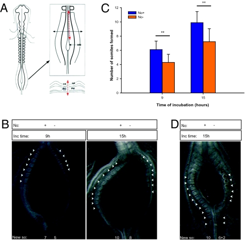
Fig. 2.
Notochord ablation in ovo delays somite segmentation of the undetermined PSM. (A) Schematic representation of embryo microsurgical operation. Only one of the embryo's PSMs (PM) was left in contact with the notochord (No). HN, Hensen's node; FP, floor plate; NP, neural plate. (B) Representative results obtained in operated embryos after a reincubation period of 9 or 15 h. (C) Graphic representation of the mean number of somites formed after 9 or 15 h. Data are mean ± SD. **P < 0.001. (D) Operated embryo reincubated for 15 h where a gap in somite formation is observed, resulting from the absence of notochord (No−) in the PSM tissue juxtaposed to the surgical slit, whereas reestablished contact of the posterior PSM with the notochord reinstates timely somite formation. Presence (No+) or absence (No−) of notochord is indicated. Black arrowheads indicate somites formed before the surgical intervention; white arrowheads indicate somites formed during reincubation (New so).

