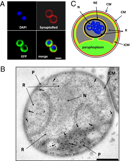
Fig. 2.
Internalized proteins in G. obscuriglobus cells are localized to paryphoplasm. (A) G. obscuriglobus cells were incubated with GFP and then stained with DAPI and SynaptoRed. A GFP-containing region is seen in the cytoplasm bounded by the cytoplasmic membrane as defined by the SynaptoRed staining and is separated from the nuclear body (DAPI staining). (B) TEM image of a section of high-pressure frozen cryosubstituted cells of G. obscuriglobus, immunogold-labeled to detect GFP via anti-GFP antibody and secondary antibody conjugated with 10 nm colloidal gold. Gold particles (short arrows) labeling internalized GFP are only associated with paryphoplasm (P). Gold particles were excluded from both the double membrane-bounded nucleoid and the riboplasm. The riboplasm (R), fibrillar nucleoid (N), and intracytoplasmic membrane (ICM) are indicated. (Scale bar, 500 nm.) (C) Diagram representing the functional compartmentalization of G. obscuriglobus. N, nucleoid; NE, nuclear envelope; ICM, intracytoplasmic membrane; R, riboplasm; CM, cytoplasmic membrane; CW, cell wall.

