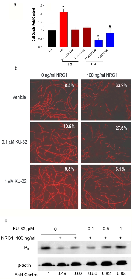
Figure 2. KU-32 protects rat sensory neurons against glucose-induced death and neuregulin-induced demyelination.
(a) Embryonic sensory neurons were treated for 6 h with 1% DMSO or 0.1–1 μM KU-32 in a medium containing 25 mM glucose (LG). The glucose concentration was raised to 45 mM to induce hyperglycaemia (HG) and the cells were incubated for an additional 24 h. Cell death was assessed using calcein AM and propidium iodide. *P< 0.05 versus LG control; ∧P<0.003 versus HG; #P<0.02 versus HG (n = 3). (b) Myelinated rat SC-DRG neuron co-cultures were treated overnight with 1% DMSO, 0.1 or 1 μM KU-32 and the cultures treated with PBS or 100 ng/ml NRG1 for 48 h. The myelin segments were visualized by staining for MBP. Numbers show percentage degenerated segments in each culture. Results are from a single experiment performed twice with similar outcomes. (c) Myelinated rat SC-DRG neuron co-cultures were treated as above and immunoblot analysis for the P0 compact myelin protein was performed. Band intensities were normalized to β-actin and expressed as a percentage of the control. Results are from one experiment performed twice with similar outcomes.

