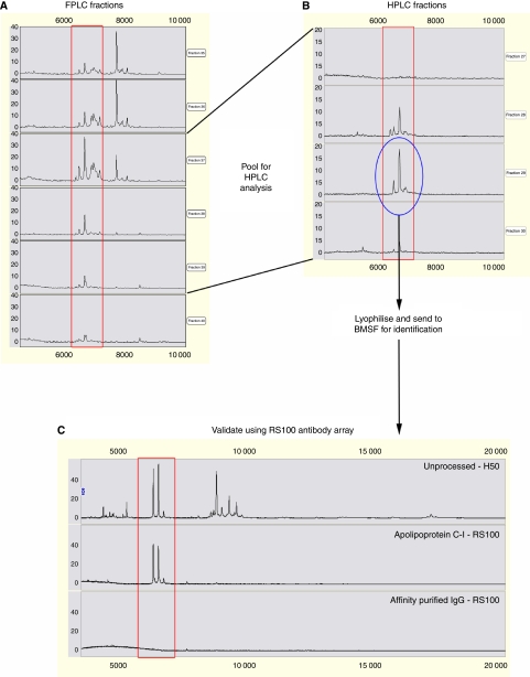
Figure 2.
Purification, identification and validation of the apparent 6.6 kDa proteins from the serum of a patient with pancreatic adenocarcinoma. (A) Proteins from serum were run through a Superose 12 HR 10/300 GL column, eluted with 0.1 M acetic acid/0.1 M NaCl (pH 3.0) and fractions were monitored by SELDI-TOF MS using NP20 chips. (B) Pooled fractions were further purified by reverse-phase HPLC with a 40 min 15–60% acetonitrile gradient in 0.1% trifluoroacetic acid and fractions were monitored by SELDI-TOF MS using NP20 chips. The fraction containing the 6.6 kDa proteins (circled) was then lyophilised and sent to BMSF for identification. (C) Identification of the 6.6 kDa proteins was validated using a SELDI immunoassay approach (RS100 antibody array).

