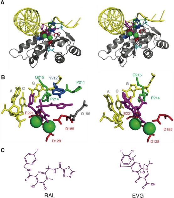
Figure 5.
Crystal structures of PFV IN in complex with INSTI. A. Three-dimensional structure of the PFV IN core domain in complex with its viral DNA substrate and RAL (left) or EVG (right) in the presence of magnesium. The core domain (amino acids 123–269) is colored in grey. Positions 212, 217 and 224, corresponding positions 143, 148 and 155 in HIV-1 IN are highlighted in blue. Positions 161 and 209, corresponding to secondary mutation positions 92 and 140 for HIV-1 IN, are highlighted in cyan. Cartoon representations were obtained using MacPyMol version 0.99rc6 and the pdb files 3L2T (RAL) and 3L2U (EVG) containing the PFV IN complete structure with viral DNA and Mg2+/Zn2+. B. Close view of the active site containing RAL (left) or EVG (right). Only the residues of PFV IN and the bases of the viral DNA within 5 Å of the drug are represented as sticks. Colors are similar to panel A. C. RAL (left) and EVG (right) chemical structures oriented as in the PFV IN structure in panel B.

