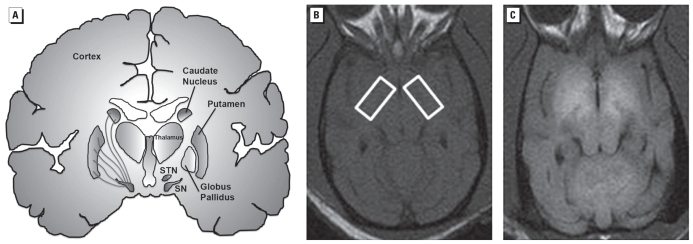
Figure 1.
(A) Schematic depicting the different brain structures comprising the basal ganglia (labeled in the right hemisphere). The left hemisphere shows the nigrostriatal dopamine fibers whose cell bodies are located in the substantia nigra (SN) and innervate the caudate and putamen. These are the axonal projections that degenerate in Parksinson’s disease. T1-weighted MRI at the level of the globus pallidus of a control nonhuman primate brain (B; boxed areas) and a nonhuman primate brain exposed to Mn (C). Note the increase in signal intensity (white areas) in the Mn-exposed animal (C) relative to the control animal (B). SNT, subthalamic nucleus.

