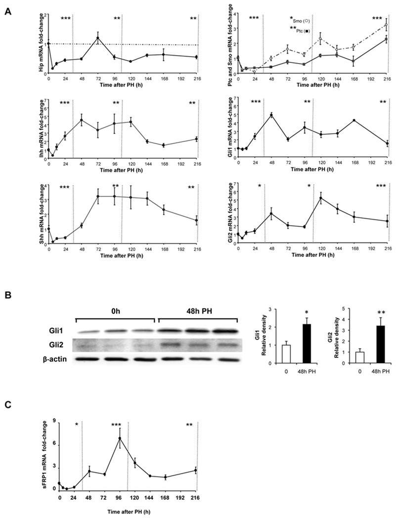
Figure 1. Activation of the Hedgehog pathway after partial hepatectomy (PH).
PH was performed on 102 mice; 6–12 mice/group were sacrificed at 6h, 12h, 24h, 48h, 72h, 96h, 120h, 144h, 168h and 216h post-PH. Quiescent (0h) livers and regenerated livers were harvested and total RNA from each mouse was examined in triplicate by qRT-PCR for (A) Hh pathway signaling components, Hip, Ihh, Shh, Ptc/Smo, Gli1, and Gli2 and (C) sFRP1, a Hh-target gene. Results are expressed as fold change relative to 0h liver tissues; mean ± SEM are graphed. Data are grouped into pre-replicative, replicative and post-replicative sets for statistical analysis using one-way ANOVA. (B) Representative Western blot analysis of Gli protein expression in 0h and post-PH livers of 3 randomly selected mice that were sacrificed at 48h post-PH. β-actin was used as a loading control; blots were densitized; Gli expression was normalized to β-actin expression in each sample; cumulative data were graphed. *P<0.05 or **P<0.01 or ***P<0.001 vs 0h.

