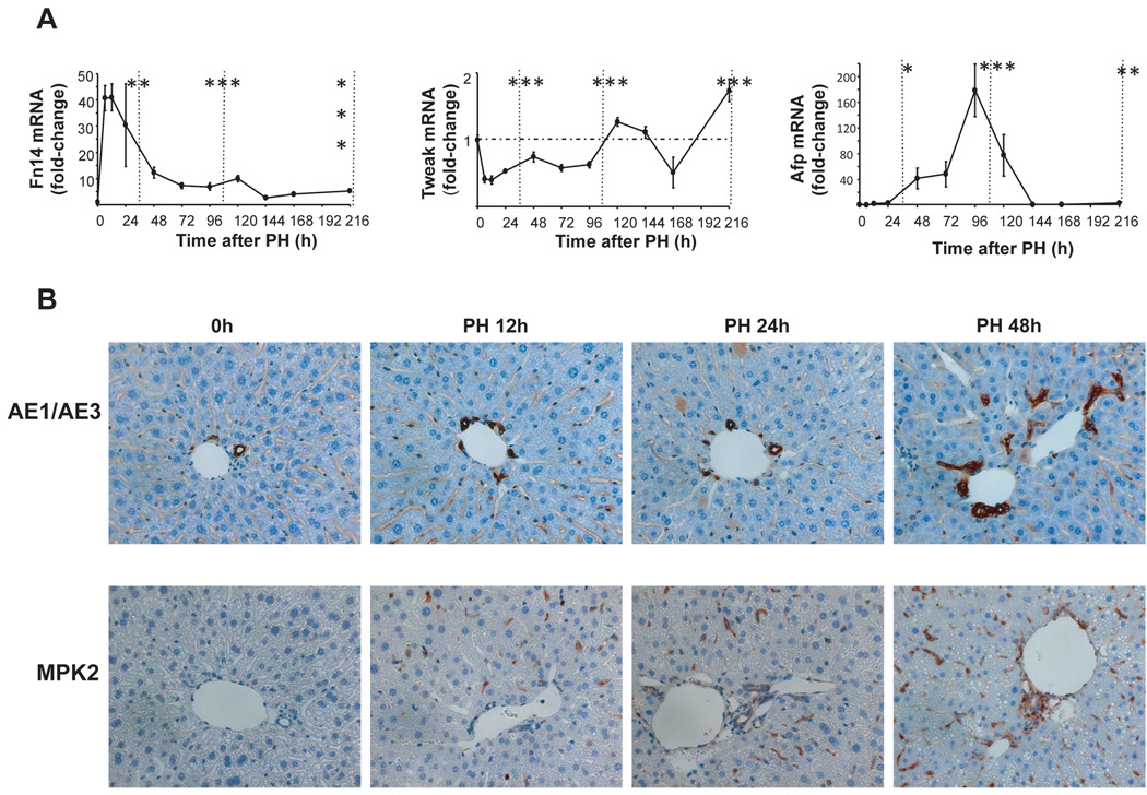
Figure 2. Changes in markers of liver epithelial progenitors after PH.
Effects of PH on mRNA levels of progenitor markers were examined in all 102 mice described in Figure 1. (A) QRT-PCR analysis of Fn14, Tweak, Afp. Results are expressed as fold change relative to 0h liver tissues; mean ± SEM are graphed. Data are grouped into pre-replicative, replicative and post-replicative sets for statistical analysis. *P<0.05 or **P<0.01 or ***P<0.001 relative to time 0h (ANOVA). (B) Two other progenitor markers, AE1/AE3 and Mpk2, were evaluated using immunohistochemistry in 0h and post-PH livers from randomly selected mice that were sacrificed at either 12h, 24h or 48h post-PH (n=4 mice/time point). Representative sections are displayed. AE/AE3 or Mpk2-stained cells are brown.

