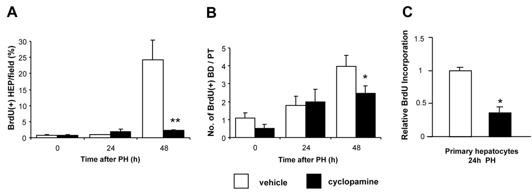
Figure 6. Inhibition of Hedgehog-signaling impairs proliferation of hepatocytic and ductular cells after PH.
(A) Percentage BrdU(+) hepatocytes per field and (B) number of BrdU(+) ductular cells per portal tract in liver sections from all surviving cyclopamine- or vehicle-treated mice described in Fig. 5. *P<0.05, **P<0.01 vs comparable vehicle-treated control (Student’s t-test). (C) Direct effects of cyclopamine on proliferative activity of primary hepatocytes. Two additional pairs of mice were subjected to either sham surgery or PH; hepatocytes were isolated 24h later and cultured overnight in the presence of BrdU ± either tomatidine (an inactive cyclopamine analog) or cyclopamine (5 µM); for each treatment group BrdU incorporation was assessed in quadruplicate plates by ELISA. Mean ± SEM BrdU incorporation in cyclopamine-treated cultures was normalized to results in respective tomatidine-treated cultures. *P<0.05 vs comparable tomatidine-treated controls (Student’s t-test). Cyclopamine had no effect on BrdU incorporation in hepatocytes from sham-operated mice (data not shown).

