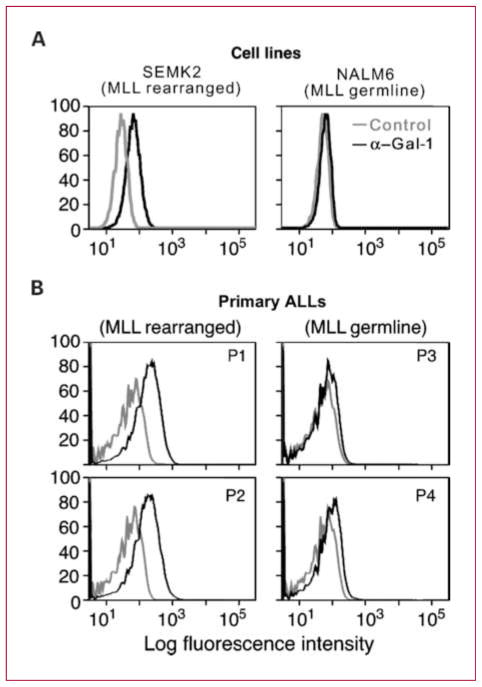
Fig. 2.
Gal-1 is detected by intracellular flow cytometry in MLL-rearranged B-ALL cell lines and primary tumors. Intracellular flow cytometry was performed on B-ALL cell lines (A) and viable primary tumor specimens from four B-ALL patients with known MLL translocation status (B). B, mean fluorescence intensity for control and anti–Gal-1 immunostaining was as follows: P1, 21.5 versus 108; P2, 24.2 versus 117; P3, 20.3 versus 35.5; and P4, 26.6 versus 63.3.

