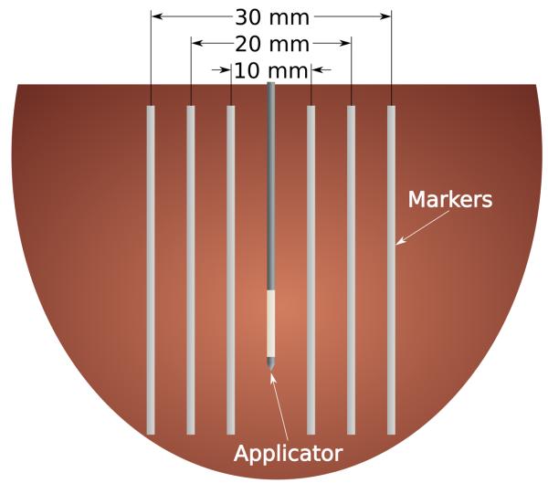
Figure 1.
Cartoon section of the experimental setup. Thin, flexible markers were placed on either side of the ablation applicator to mark the original position of the tissue at peripheral (30 mm), middle (20 mm) and inner (10 mm) locations. Lung samples only included the peripheral and inner points due to the large amount of contraction that occurred during ablation.

