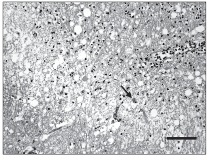
Figure 2.
Microphotograph of the left thalamus (index horse). This is the junction between the necrotic zone (up right) and surrounding thalamus (down left). There is intense vacuolization of the neuropil and infiltration by numerous neutrophils in the necrotic area. Note a necrotic neuron (arrow). Hematoxylin-eosin-saffron (HES). Bar = 50 μm.

