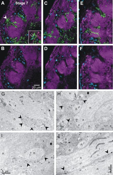
Figure 8.
Identity of MasGAT-positive processes. A-B, C-D, E-F. Glial nuclei in blue. Upper panels (A,C,E) show a portion of stage-7 ALs double-labeled single optical sections with anti-MasGAT (green) and anti-HRP (magenta); lower panels (B,D,F) show only anti-HRP labeling. Comparison of upper and lower panels reveals no overlap between MasGAT-positive and HRP-positive processes, except possibly at the distal tips of the MasGAT-positive processes. These processes are extremely thin so that glial and neuronal processes can occupy the same Z volume. Inset: The labeled processes in A were traced to the cell body that appeared in the position in the glial border indicated by the arrowhead but that was obscured by overlying labeled processes in the full 3-D reconstruction. G-J. Electron micrographs taken in glomerular neuropil with MasGAT indicated by the presence of silver-enhanced gold particles. Some (arrowheads), but not all (diamonds), glial processes contain gold particles. Neuronal processes (n) have no labeling above background.

