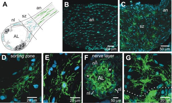
Figure 9.
MasGAT in the developing nerve and nerve layer of the antennal lobe from a stage-7 animal. A. Schematic diagram of the M. sexta nerve and antennal lobe. Peripheral glial cells populate most of the length of the antennal nerve (an), migrating from the antennal sensory epithelium. Centrally derived glial cells populate the sorting zone region (sz), the nerve layer (nl), and form the glomerular borders. B. No MasGAT-positive glial cells were found among the peripheral glial cells. A subset of glial cells in the SZ (C-E) and in the nerve layer (F-G) were MasGAT-positive during the period of ORN axon ingrowth. D-E and G show higher power views of the morphology of individual MasGAT-positive glial cells. Dotted line in G indicates the border between the nerve layer, above, and the apical edge of a glomerulus, below. *, glomerulus.

