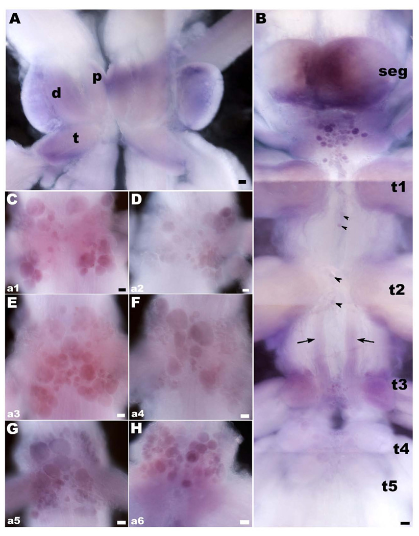
Figure 3. OA/TAMac receptor’s mRNA in the CNS of the prawn.
A: Brain OA/TAMac receptor mRNA appears as diffuse probe labeling in the neuropil areas of the protocerebrum (p), deutocerebrum (d), and tritocerebrum (t). B: Between the subesophageal ganglion (seg) and the first thoracic ganglia (t1), there is a central cluster of cell bodies of varying sizes that showed OA/TAMac mRNA staining. In the thoracic ganglia, two to four stained cell bodies showed mRNA labeling along the ventral midline (black arrowheads), as did a group of cells in the ventral middle region of the t3–t5 ganglia. There is also diffuse staining in a bilateral bundle of processes extending from t2 to t3 (black arrows). C–H: OA/TAMac mRNA Staining was also observed in all abdominal ganglia. Cells are arranged in clusters, following a pattern that repeated itself in each of the six ganglia (a1–a6). The pattern consisted of cells located ventrally at the center of each hemiganglion, along with 4 clusters of cells, arranged as the wings of a butterfly, located towards the dorsal lateral-most edges of each ganglion. Scale bar = 100 µm.

