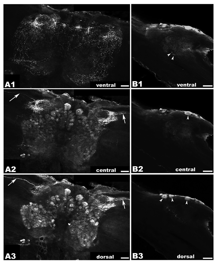
Figure 4. OA/TAMac immunoreactivity (ir) in the brain and circumesophageal (CEG) ganglia.
A1–A3: Brain OA/TAMac-ir was observed as punctate staining in the ventral part of the ganglion (A1), in the protocerebrum, lateralmost aspects of the deutocerebrum, and extending towards the inferior aspects of the tritocerebrum. Moving more deeply towards the center of the ganglion (A2), punctate staining is observed in the lateralmost protocerebrum, while medium- and large-sized cells are found more medially. Highly immunoreactive structures within the nucleus, possibly nucleoli, are observed in all stained cells. Bilateral axons are observed in the optic nerves going towards a region of intense punctate staining in the lateral protocerebrum (white arrows). More dorsally (A3), protocerebrum punctate staining and the bilateral axons (white arrows) are still visible. Smaller-sized cell bodies showing OA/TAMac-ir are also observed in the deutocerebrum. B1–B3: In circumesophageal ganglia, OA/TAMac-ir was observed as punctate staining towards the center of the ventral (B1), middle (B2), and dorsal (B3) aspects of the ganglia. OA/TAMac-ir is also observed in two small cells in the ventral ganglia (white arrowheads; B1), and in one to three medium-sized cells on the outer surface of the ganglia (white arrowheads; B2–B3). Scale bar = 100 µm.

