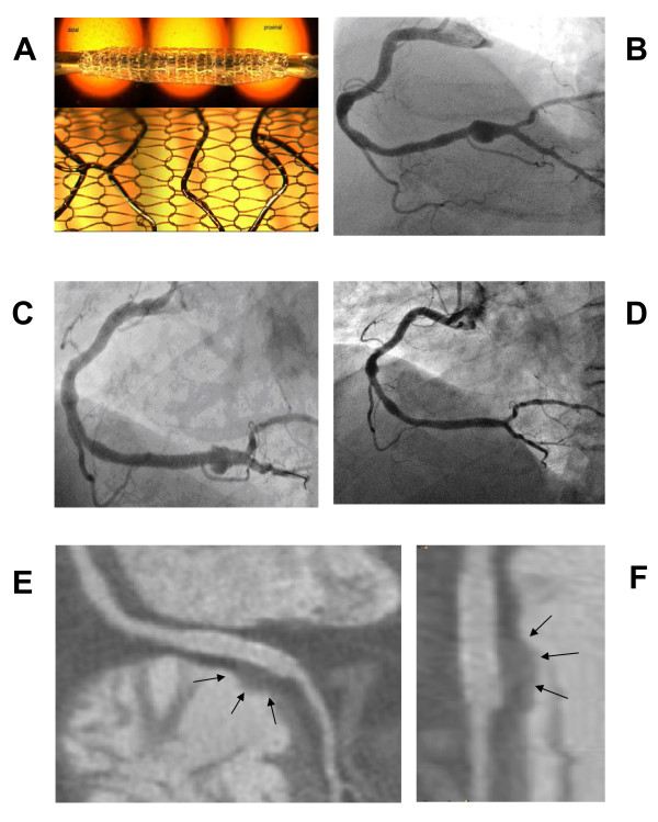
Figure 1.
Coronary angiography and CT scan before and after MGuard stent implantaion. (A) The unexpanded (top) and the expanded stent covered with a sleeve mesh (bottom). Reproduced with permission from InspireMD. (B) Angiography of the right coronary artery with the presence of a large saccular aneurysm involving the distal part of the artery to the crux cordis. (C) Partial opacification of the aneurysmal sac through the holes of the mesh just after stent implantation. (D) Coronary angiography at one month follow-up showing the exclusion of the aneurysm. (E) Coronary CT scan at one month: multiplanar reformation of the right coronary artery near the crux cordis; on the right ventricle side of the distal part of the stent, is clearly demonstrated the water density remnant of the treated aneurysm (arrows): low density fat is surrounding the proximal stent. (F) Coronary CT scan at one month: The magnified view of the stent allows for a better identification of the treated aneurysm (arrows).

