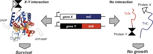
Fig. 1.
Schematic of an AK-based PCA. TnN (red) and TnC (blue) fragments are engineered to only complement thermophile growth when they are fused to proteins (X and Y) that drive their association. The AKTn fragments are mapped onto the structure of an AK ortholog (PDB ID, 1zin) (Bae and Phillips, 2004) with bound P1,P5-di(adenosine-5) pentaphosphate (peach) and zinc (gray) shown as space filling models. The side chains of the cysteines mutated in AKTn (C133 and C156) are shown in green.

