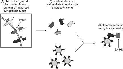
Fig. 4.
Schematic of strategy for classifying scFvs capable of binding extracellular epitopes using tryptic fragments derived from intact cells. In Step 1, biotinylated plasma membrane proteins are digested with trypsin to release extracellular fragments. The cleaved fragments are incubated with an isolated scFv displaying yeast in Step 2. In Step 3, the interaction between extracellular fragment and scFv is quantified using a flow cytometer. If binding is retained using the tryptic fragments, an scFv is classified as binding to an extracellular epitope (Table I).

