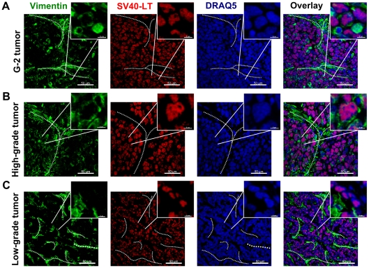
Figure 4. Expression of the intermediate filament protein vimentin and the SV40-LT transgene in G-2 and WAP-T tumors.
(A–C) Representative confocal images of tumor-cryosections of a G-2 cell-derived tumor (A), a high-grade (B) and a low-grade (C) endogenous WAP-T tumor stained with anti-vimentin (green) and anti-SV40-LT (red) antibodies. The nuclei were stained with DRAQ5 (blue). The power insets are used to display co-expression of vimentin and SV40-LT in G-2 and high-grade WAP-T tumors. The white dashed lines mark stromal structures. The confocal 3D-stacks were deconvoluted using Huygens Essential software and reconstructed with the Imaris software. Scale bar: main picture: 50 µm; magnification: 6 µm.

