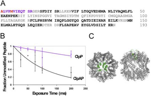
FIGURE 1. Synchrotron hydroxyl radical footprinting results.

A) Tryptic peptide coverage of ClpP. Color coding of amino acids: Black: MSMS identified peptides, Blue: peptides with modification, Red: specifically identified modified residues, Grey: not identifiable in spectra. B) ClpP peptide 1–12 oxidation curves. blue: ClpP tetradecamer, black: ClpAP complex. C) Left: ClpP top view, Right: ClpP side view. N-terminal residues are highlighted in green. Structure is from Bewley et al. (20), PDB accession code 1YG6.
