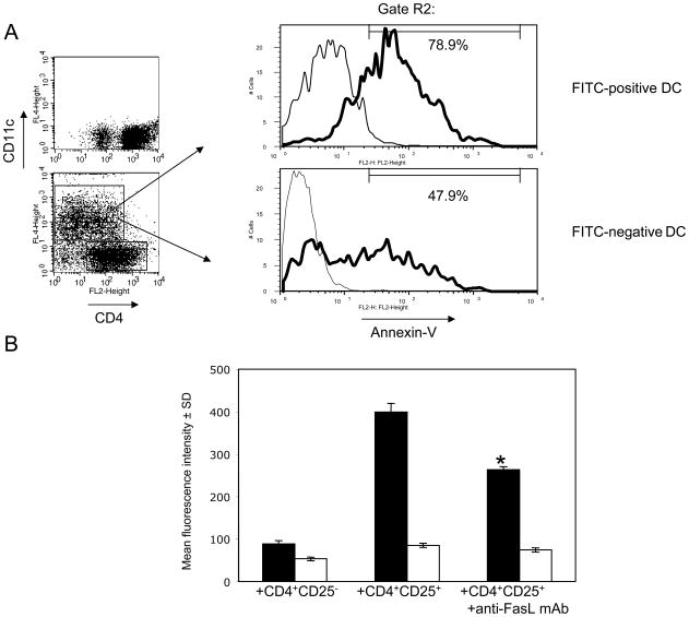
Figure 3. CD4+CD25+ T cells kill FITC+, but not FITC− DC through Fas-FasL interactions during co-culture.
(A)2 × 10⁵ cell aliquots of purified CD4+CD25+ T cells were co-cultured in triplicate with 105 DC purified from skin-draining lymph nodes of FITC-sensitized mice. After 4 h of culture, cells were stained with APC-labeled anti-CD11c mAb and with Annexin-V-PE. Dot plots show control aliquots of CD4+ T cells cultured alone (upper panel) or with DC (lower panel) and stained with anti-CD4 and anti-CD11c mAb. CD11c-positive cells were gated (gate R2) and then FITC+ or FITC− DC within this gated population were analyzed by histogram for Annexin-V staining (solid histograms). The numbers in histograms indicate the percentages of Annexin-V+ DC depicted by the gate that excludes autofluorescent DC that were not stained with Annexin-V (thin histograms). The experiments were repeated three times with similar results.
(B)Purified CD4+CD25+ T cells or CD4+CD25− T cells were cultured in triplicate with DC purified from FITC-sensitized mice for 4 h, and then stained with APC-labeled anti-CD11c mAb and with Annexin-V-PE. FITC+ DC (■) and FITC − DC (□) were gated as described in Figure 2A and analyzed by histograms for the fluorescence intensity of Annexin-V staining. Purified anti-FasL mAb was added to CD4+CD25+ T cells and DC cultures at 25 ug/ml. * P < 0.05 as compared to CD4+CD25+ T cells and DC co-cultures without anti-FasL mAb. The experiments were repeated two times with similar results.

