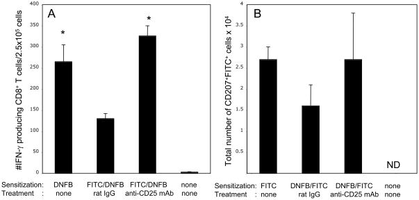
Figure 6. CD4+CD25+ T cells can suppress activation of hapten-specific CD8+ T cells in a non-specific manner.
(A) Mice were sensitized with 1% FITC on day 0 and were given i. p. injections of 250 μg of control rat IgG or anti-CD25 mAb on days 0, +1 and +2. On days +5 and +6 post-sensitization mice were sensitized with 0.25% DNFB. Five days after DNFB sensitization, CD8+ T cells enriched from LNC of sensitized mice were cultured with DNBS-labeled splenocytes and the numbers of DNFB-specific IFN-γ producing CD8+ T cells were evaluated by ELISPOT. Mice sensitized with DNFB without FITC pre-sensitization and non-sensitized mice were used as positive and negative controls. *P < 0.01 by two-tailed Students’ t test as compared to the group of mice sensitized with FITC and DNFB and treated with rat IgG.
(B) Mice were sensitized with 0.25% DNFB on days 0 and +1 and were given i. p. injections of 250 μg of control rat IgG or anti-CD25 mAb on days 0, +1 and +2. On day +5 post-sensitization mice were sensitized with 1% FITC. On day +3 after FITC sensitization, LNC suspensions were stained with PE-labeled anti-CD11c mAb and AlexaFluor647-labeled anti-CD207 mAb. FITC-presenting LC were identified as CD207+FITC+ cells within the CD11c+ population. Total numbers of CD207+FITC+ cells in the skin-draining lymph nodes of each individual mouse were calculated based on the percentage of these cells in each analyzed cell aliquot. ND – not detected.

