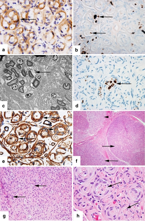
Figure 3.
This set of images shows our pathologic specimen of intraneural perineurioma with appropriate staining methods. a Epithelial membrane antigen. Original magnification ×1,000. Epithelial membrane antigen (EMA) positive staining shows pseudo-onion bulbs consisting of perineurial cells (arrow). b S-100 stain. Original magnification ×1,000. S-100 staining highlights scattered Schwann cells within the fascicles (arrow). This again shows that the whorls are not composed of Schwann cells and therefore are pseudo-onion bulbs. c Electron microscopy. Direct magnification ×2,500. Electron microscopy shows perineurial cells circumferentially enwrapping multiple axons (arrow). d Neurofilament stain. Original magnification ×1,000. Neurofilament staining highlights scattered axons within the fascicles, some of which are in clusters (arrow) surrounded by perineurial cells. In chronic lesions such as this, axons are scant or absent. e Collagen IV stain. Original magnification ×1,000. Collagen IV highlights perineurial cells’ basement membranes (arrow). High quantities of collagen deposition as seen in this image occur with long-standing lesions. f Hematoxylin and eosin stain. Original magnification ×100. A low power cross-section view of the affected nerve showing involved fascicles (arrows) and a relatively uninvolved fascicle (arrowhead). Involved fascicles are enlarged and hyperchromatic. g Hematoxylin and eosin stain. Original magnification ×400. Pseudo-onion bulbs (arrows) consisting of multilayer cuffs of perineurial cells surrounding single or multiple nerve fibers, seen at higher magnification in c. h Hematoxylin and eosin stain. Original magnification ×1,000. High power view showing large pseudo-onion bulbs surrounding multiple axons (arrow).

