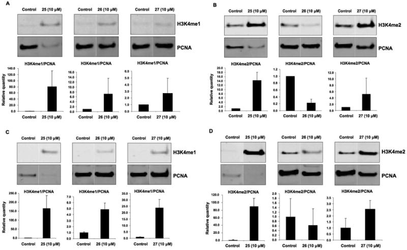
Figure 3.

Effect of compounds 25-27 on the expression of global H3K4me1 and H3K4me2. Calu-6 human anaplastic non-small cell lung carcinoma cells were treated with a 10 μM concentration of 25, 26 or 27 for 24 h (panel A and B) or 48 h (panel C and D) as described in the methods section. Panel A and C shows global H3K4me1 expression and panel B and D shows global H3K4me2 expression. Proliferating cell nuclear antigen (PCNA) was used as a loading control. Shown are Western blot images from a single representative experiment performed in triplicate. Relative protein expression levels were determined by quantitative Western analysis using the Odyssey infrared detection system shown as bar graphs. The results represent the mean of three treatments ± SD. The protein expression level for control samples was set to a value of 1.
