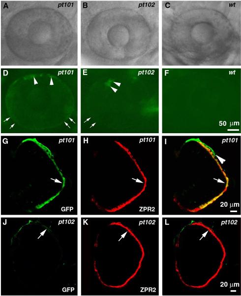
Figure 3.
The Fugu tyrp1 promoter directs GFP expression in RPE cells in pt101 and pt102. A—F. The lateral views of the eyes of pt101, pt102, and wildtype under a fluorescence stereomicroscope at 72 hpf. A—C are images taken in bright field and D—F are their respectively corresponding images in GFP fluorescence field. Arrows indicate the stronger GFP appearance at the boundary regions of the eyes. Arrowheads indicate what is likely to be choroidal melanophores adjacent to the RPE. G—I. In pt101, GFP signal in the eye (G, arrow) colocalizes with RPE specific marker zpr2 (H, arrow) at 5 dpf. I is the merged image of G and H. The arrowhead in I indicates the choroidal melanophores. J—L. In pt102, the weak GFP expression (J, arrow) in the eye also localizes to the zpr2-stained RPE (K). L is the merged image of J and K.

