
Figure 4.
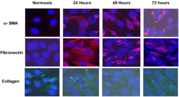
Immunostaining performed for HIF-1α, α-SMA, collagen, and fibronectin in normoxic and hypoxic fibroblasts. Cells staining for red are positive for α-SMA and fibronectin and those staining green are positive for collagen. This demonstrated that there was increased expression of α-SMA, fibronectin, and collagen over time suggesting that fibroblasts were converting their phenotype to myofibroblasts under hypoxia.