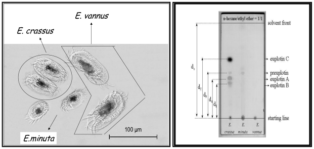
Figure 2.
Left: Transmission electron microscopy (TEM) image of Euplotes crassus, Euplotes minuta and Euplotes vannus ciliates; Right: TLC plate (stationary phase: Merck Si60 PF254, eluent: n-hexane-ethyl ether 1:1, staining reagent: Ce(SO4)2/H2SO4) of crude cell extracts (0.1 mL of cell pellet) from typical strains of E. crassus, E. minuta and E. vannus.

