
Figure 2.
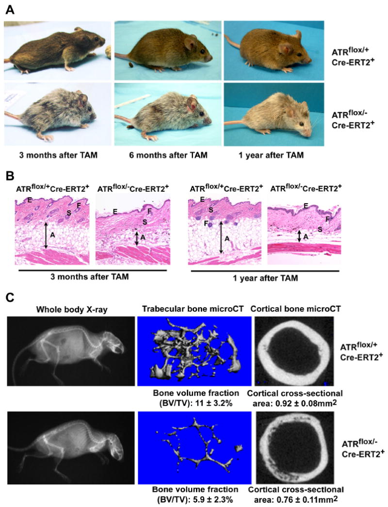
ATR deletion leads to hair graying, alopecia, kyphosis and osteoporosis. (A) ATR deletion following TAM treatment leads to pervasive hair graying and patchy hair loss and kyphosis in ATRmKO mice. (B) Age-related abnormalities in the skin of ATRmKO mice. Thinning of the subcutaneous adipose layer (A), thickening of the epidermis (E), loss of hair follicles (F) and sebaceous gland cell hypertrophy (S) were observed in ATRmKO mice (n = 19) but not in control mice (n = 22). Sections from sex-matched mice are shown. (C) Increased kyphosis and osteoporosis in ATRmKO mice. Control and ATRmKO mice were X-rayed 1 year after TAM treatment (left panel). Increased kyphosis over controls was observed in all ATRmKO mice analyzed (n = 6). To analyze bone volume and cross-sectional area, femurs from ATRmKO and control mice were subjected to microCT analysis (4-7 mice analyzed/group). Trabecular bone in the distal metaphysic (middle panel) and cortical bone cross-sectional area (right panel) was imaged and analyzed. Bone volume/total volume (BV/TV) and cortical area are shown as mean ± SD. P ≤ 0.04 as calculated by Student's T-test.