
Figure 4.
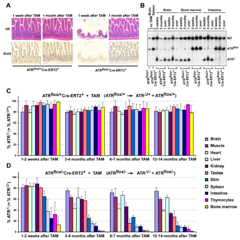
ATR deletion leads to loss of proliferating cells in mice. (A) Rapid loss and reconstitution of proliferating intestinal epithelial cells after ATR deletion. Mice were treated with BrdU in drinking water for up to 1 month following TAM treatment. Loss of proliferating intestinal epithelial cells was observed 1 week after ATR deletion in ATRmKO mice (n = 4), however, a full recovery was observed 3 weeks later (n = 5). Pictures were taken at 100× magnification. (B) ATRΔ/- cells are rapidly lost in proliferating tissues (bone marrow, intestine), but not in the brain of ATR knockout mice. Southern blot of genomic DNA isolated from ATRflox/+Cre-ERT2+ (control) and ATRflox/-Cre-ERT2+ (ATRmKO) mice treated or left untreated with TAM. Appearance of the ATRΔ allele represents ATRΔ/+ cells in control mice and ATRΔ/- cells in test mice. (C, D) Percentage of ATRΔ/+ cells remains constant (C), while ATRΔ/- cells are lost (D) in various tissues following lox recombination. Southern blot band intensities of lox recombined (ATRΔ) and unrecombined (ATRflox) were used to quantify the total percentage of cells that maintained a recombined copy of ATR (ATRΔ). Band intensities were quantified by phosphoimager, and mean percentages were calculated from the ratio ATRΔ over ATRΔ + ATRflox. For each tissue and time point, 2-8 mice were evaluated; each error bar indicates one standard deviation.