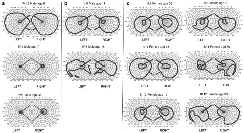
Figure 4.
(a) Top panel shows the visual field OD from IV:13 at 6 years of age. The visual field for IV-4-e is full. In contrast, the middle panel shows the visual field OD from IV: 1 at 7 years of age. Visual fields for both IV-4-e and I-4-e stimuli are severely restricted. Although virtually all males show a severely constricted I-e visual field between 10° and 20° in maximum diameter, there is variability in the initial visual field deficit for the IV-4-e isopter. The bottom panel shows visual fields of patient IV: 1 at the age of 22 years. Despite considerable constriction of visual fields in this patient from a very young age, his central vision remained preserved into early adulthood. (b) The top panel shows visual fields for the V-4-e and II-4-e isopters from family member IV: 6 at 11 years of age. There is superior restriction of fields bilaterally. At 11 years of age, the patient was unable to visualise the I-4-e isopter, hence the II-4-e isopter is shown in the top panel. Even the II-4-e stimulus visual field was significantly contracted to 20° full diameter. The bottom panels show visual fields from the same individual at 15 years of age when the patient could visualise the I-4-e isopter, but as with other males, that field was severely constricted. In addition, a significant loss to the inferior visual field (V-4-e) had occurred bilaterally. (c) Each panel shows the visual field IV-4-e and I-4-e from a female family member at initial evaluation and at follow-up. I-4-e fields are markedly reduced, although they are still larger than those in affected males. IV-4-e fields are constricted to a lesser degree but morphologically normal. There are varying degrees of regional scotomata in the IV-4-e isopter field on follow-up. I-4-e isopter fields are globally constricted.

