
Figure 3.
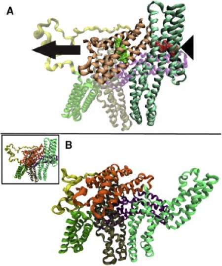
Simulation of vinculin activation by simultaneous interaction. To simulate the stretching of vinculin due to simultaneous interaction with actin and talin, force was applied to Vt (binds to actin) away from the VBS binding region of Vh. (A) The direction of force is indicated by the arrow. The four residues nearest the center of mass of the VBS binding region of Vh were constrained (triangle) while 50 pN and 100 pN of constant force was applied to the four residues nearest the center of mass of Vt (residues shown in wire mesh). (B) After >300 ps of simulation, the VBS binding region of Vh moved away from Vt. This conformational change involved bending of the helical regions in domain 1. As the simulation progressed, the VBS binding region moved farther away from Vt. Shown here is the conformation of vinculin with the VBS binding region ∼120 Å away. In this conformation, the steric hindrance preventing Vt interaction with actin is removed and vinculin is likely activated.