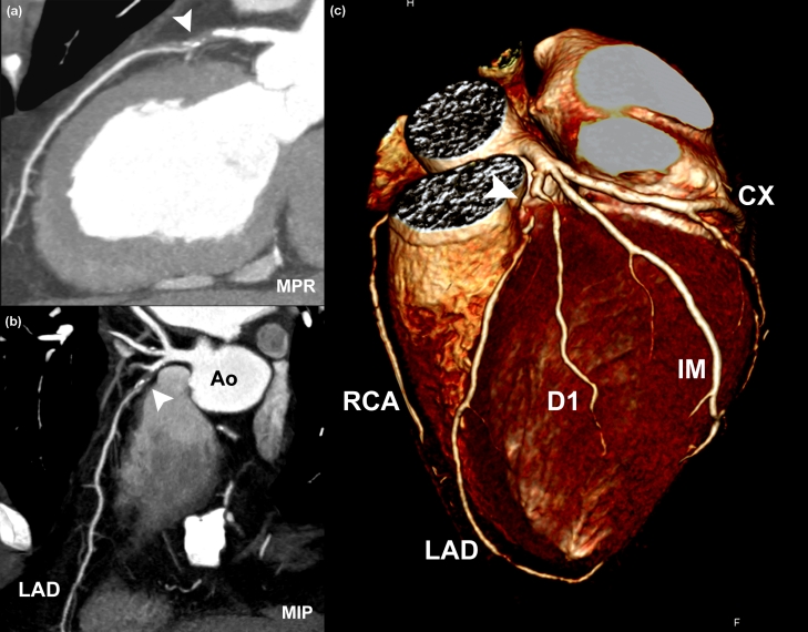
Figure 2. Example of retrospectively gated dual-source CT coronary angiography.
Heart rate 64 beats/minute; pitch 0.28; tube voltage 120 kV; peak tube current 625 mA/tube; and ECG tube current modulation. (a) The curved multiplanar projection (MPR), (b) the maximum intensity projection (MIP) and (c) the colored-volume-rendered image show a subtotal occlusion (marked by a white arrowhead) of the proximal left anterior descending (LAD) artery. Effective dose, 7.2 mSv. Ao, aorta; CX, left circumflex artery; D1, first diagonal; IM, intermediate branch; RCA, right coronary artery.

