
Figure 1.
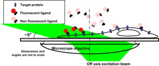
Schematics of the experimental setup. A low concentration of fluorescent ligands is introduced in the extracellular medium such that a constant rate of membrane molecules is being labeled during the imaging sequence. Oblique illumination of the sample is used to excite predominantly fluorescent ligands that have bound to the cell surface while not illuminating the molecules in the above solution.