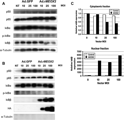
Figure 1.
MEOX2 expression results in the nuclear accumulation of p65 and IκBβ. Cytoplasmic and nuclear fractions were isolated from HUVECs transduced with either Ad.rMEOX2 or Ad.GFP (control) and subjected to western blot as described in Section 2. (A) Cytoplasmic fraction. IκBβ levels decrease slightly in response to MEOX2 expression, whereas p65 levels remain nearly constant. (B) Nuclear fraction. Nuclear IκBβ levels increase markedly in response to increasing expression of MEOX2 driven by Ad.rMEOX2. Nuclear p65 increases more modestly. No α-tubulin was detectable in the nuclear extract, indicating a lack of contamination with cytoplasmic extract, and MEOX2, a nuclear protein, was undetectable in the cytoplasm (data not shown). Blots were independently loaded using the same protein samples from the same experiment. (C) Densitometry for IκBβ, cytoplasmic, and nuclear fractions, from (A) and (B).

