Figure 5.
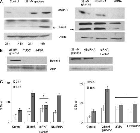
MCPIP-induced ER stress resulted in autophagy that mediated high glucose-induced H9c2 cardiomyoblast death. H9c2 cardiomyoblasts were treated with/without 28 mmol/L glucose. (A, left) At 24 and 48 h, cell lysate was collected and analysed using immunoblot with beclin-1 or LC3 antibody. β-Actin served as a control. (A, right) Cardiomyoblasts were treated with 28 mmol/L glucose with/without siRNA specific for MCPIP or with non-specific siRNA. At 48 h, cell lysate was collected and analysed using immunoblot with beclin-1 or LC3 antibody. β-Actin served as a control. (B, left) Cardiomyoblasts were treated with 28 mmol/L glucose and with 100 µM TUDC or with 50 µm 4-PBA. At 48 h, cell lysate was collected and analysed using immunoblot with antibody specific for beclin-1. β-Actin served as a control. (B, right) Cardiomyoblasts treated with 28mM glucose were treated with or without siRNA for Beclin-1 or non-specific siRNA (NS siRNA). At 48 h, cell lysate was collected and analysed using immunoblot with beclin-1 antibody. (C) Cardiomyoblasts were treated with 28 mmol/L glucose with/without siRNA specific for beclin-1 or with non-specific siRNA. At 24 and 48 h after treatment, cell death was analysed using trypan blue (left; *P < 0.05). (C, right) Cardiomyoblasts were treated with 28 mmol/L glucose and with/without 50 µM 3′MA or 1 µM LY294002. At 24 and 48 h after treatment, cell death was analysed using trypan blue (#P < 0.03).

