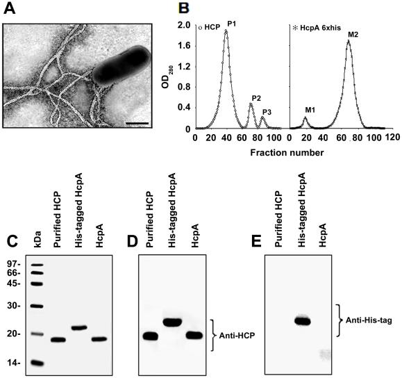
Figure 2. Immunodetection of HCP and the His-tagged HcpA protein.
Immunogold-labeling of HCP produced by EDL933 grown on Minca agar using anti-HCP antibody (A). Scale bars, 0.5 µm. HCP and the HcpA protein (without the His-tag) were purified separately using a molecular exclusion chromatography (B). Purified HCP and the recombinant protein were depolymerized in 16% SDS-PAGE gels and stained with Coomassie blue (C). Peak P1, is the highest protein peak that showed a protein of 19 kDa corresponding to the pilus subunit HcpA. Peak M2 obtained during HcpA-His purification showed a protein that migrated as a 22 kDa protein due to the presence of 6 histidines. Western blot of both proteins with anti-HCP demonstrated the presence of the pilin (D). Western blot of HcpA His-tagged protein, showing a band of 22 kDa detected with monoclonal anti-His tag antibodies (E).

