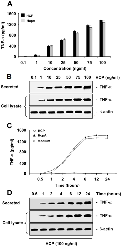
Figure 5. Induction of TNF-α release by purified HCP.
An ELISA-based assay demonstrating the induction of TNF-α in polarized HT-29 cells in response to different concentrations of purified HCP and HcpA (A). Western blot showing the kinetics of secreted proteins and cell lysates produced after the incubation of polarized HT-29 cells with HCP and HcpA (B). Both proteins stimulate the production of TNF-α in HT-29 cells in a dose-dependent manner. Purified HCP (100 pg/ml) and HcpA (100 pg/ml) were incubated with polarized HT-29 cells at different intervals (C). The supernatant recovered from the basolateral surface showed a significant time-dependent increase of TNF-α as shown by Western blotting (D). Detection of actin was used as a protein loading control.

