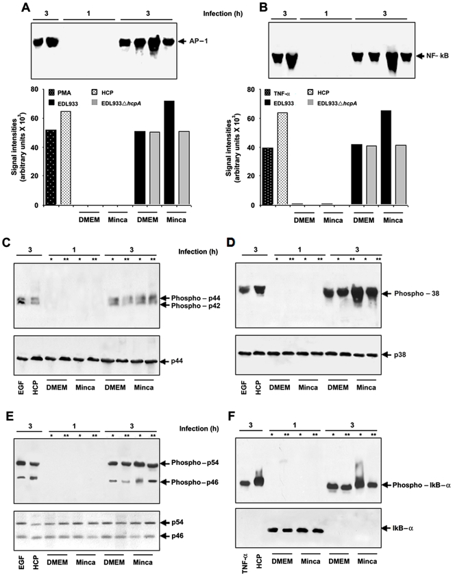
Figure 9. EHEC infection induced AP-1, NF-κB DNA binding, MAPK and IκB-α activation in HT-29 cells.
Infection with purified HCP, wild-type O157:H7 and the hcpA mutant showing percentages of AP-1 activation; PMA: Phorbol myristate acetate (A). Infection with purified HCP, wild-type O157:H7 and the hcpA mutant showing percentages of NF-κB activation (B). Western blot showing the kinetics of MAPK activation using specific antibodies that recognize the phosphorylated forms of p42 and p44 (C). Wild-type EHEC is involved in the p38 phosphorylation in conditions where HCP is produced and a significant decrease was observed when tested with the EDL933 hcpA mutant (D). ERK1/2 and JNK activation were detectable after 3 h of EHEC infection (E). IκB-α activation was detectable after 3 h of EHEC infection (F). * EDL933 O157:H7 and ** EDL933ΔhcpA.

