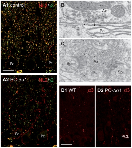
Figure 2. Synaptic organization in the cerebellum of PC-Δα1 mice.
(A1,A2) Double labelling for GABAARγ2 (green) and NL2 (red) in control (A1) and PC-Δα1 mice (A2). NL2 colocalizes extensively with the γ2 subunit and also clusters at postsynaptic sites lacking GABAARs in Purkinje cells of PC-Δα1 mice. (B,C) Electron micrographs of the ML of a PC-Δα1 mouse showing that GABA-immunopositive axon terminals (Ax) make both conventional, symmetric synapses (B, arrowheads) with Purkinje cell dendrites (Pc) and heterologous synapses (C) with spines (Sp). (D1,D2) Similar distribution of α3-GABAARs in the ML of control (D1) and PC-Δα1 mice (D2). PCL, Purkinje cell layer. Scale bars: A = 15 µm. B,C = 200 nm. D = 20 µm.

