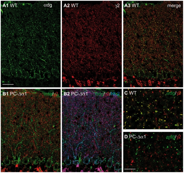
Figure 4. Dystroglycan is present at GABAergic synapses in Purkinje cells but not in cerebellar interneurons.
(A1–A3) Double labelling for α-dystroglycan (green) and GABAARγ2 (red) in the cerebellar cortex of a WT mouse. Labelling for α-dystroglycan outlines the cell bodies and major dendrites of Purkinje cells and colocalizes precisely with GABAARγ2. Labelling for the γ2 subunit is weaker at perisomatic synapses, scarcely visible in these low magnification images. (B1,B2) Triple labelling for α-dystroglycan (green), GABAARγ2 (red) and NL2 (blue) in the cerebellar cortex of a PC-Δα1 mouse. Dystroglycan colocalizes with NL2 exclusively at silent synapses that lack GABAARs (B2, cyan). NL2 associates with GABAARs at interneuron-interneuron synapses, where immunolabelling for α-dystroglycan is not visible (B2, magenta). (C,D) High-magnification images of the ML showing that in WT mice a subset of GABAergic synapses contain α-dystroglycan (yellow clusters), whereas in PC-Δα1 mice α-dystroglycan never colocalizes with GABAARγ2-positive clusters. Scale bars: A,B = 30 µm. C,D = 10 µm.

