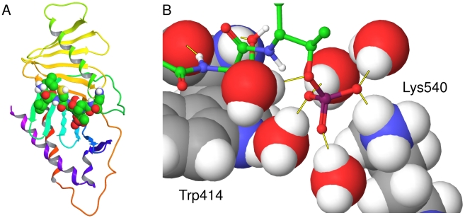
Figure 1. The PBD of PLK1 crystallised with the CDC25c phosphopeptide.
The crystal structure of the PBD of PLK1 in complex with the CDC25c phosphopeptide LLCSpTPN from PDB ID 3BZI. (a) The protein represented as a ribbon diagram with the phosphopeptide displayed by atom-colored space filling (b) The binding site shown in more detail. The phosphopeptide is displayed as atom colored ball and sticks, interfacial water molecules are displayed in atom-colored CPK and residues Trp414 and Lys540 are named in black and displayed in atom-colored CPK. Key hydrogen bonds are displayed as yellow lines.

