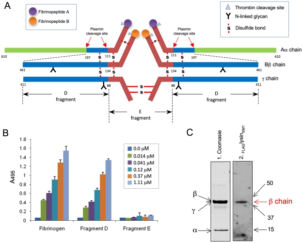
Figure 5. Binding of FLAGlysinSM1 to immobilized fibrinogen fragments D and E.
(A) Schematic diagram of human fibrinogen. The Aα, Bβ, and γ chains, major disulfide linkages, and plasmin cleavage sites are shown. (B) Indicated concentrations of FLAGlysinSM1 were incubated with immobilized human fibrinogen, fibrinogen fragment D, or fragment E. Bound FLAGlysinSM1 was detected with anti-FLAG antibody. Bars indicate the means (± S.D.). (C) Fibrinogen fragment D was separated by SDS-PAGE under reducing conditions and stained with Coomassie blue (panel 1) or transferred onto nitrocellulose membrane and incubated with FLAGlysinSM1 (5 µg/ml). The bound proteins were detected with anti-FLAG antibody (Lane 2). Arrows indicate positions of Aα, Bβ, and γ chain fragments. Numbers indicate molecular mass (kDa).

