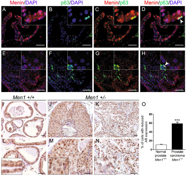
Figure 3.
Prostate cancers from Men1+/- mice do not express p63 and display heterogeneous AR expression. (A-H) Double IF staining using antibodies against menin (red) and p63 (green) performed on a normal prostate from a 24-month-old Men1+/+ mouse (A-D, n = 2) and an adenocarcinoma found in a Men1+/- mouse of the same age (E-H, n = 2). DAPI stains cell nuclei. Note that normal p63-expressing basal cells scattered within the lesion remain menin-positive (shown by white arrowheads). Boxed areas are magnified in the insets. Scale bars, 50 μm. (I-N) IHC using antibody against AR was performed on prostate tissues from Men1+/+ (I, L, n = 4) and Men1+/- (J, M and K, N, n = 4) mice. AR expression is present but more heterogeneous in an in situ prostate carcinoma (J, M, 26-month-old mouse) and an adenocarcinoma (K, N, 23-month-old mouse) from Men1+/- mice, with some cells showing greatly reduced AR expression compared to the control. Panels L-N are two-fold magnifications of the upper panels (I-K). Insets show an amplified view of a part of the prostate glands. (O) Prostatic epithelial nuclei showing absent or very low AR expression were counted in six random microscopic fields in normal prostate glands from Men1+/+ mice (n = 3) and carcinomas observed in Men1+/- prostate glands (n = 4), stained with an anti-AR antibody. The results are expressed as a percentage of total prostatic epithelial cells (250 nuclei counts). Values are means ± standard error of the mean. *** Significant difference from normal Men1+/+ prostate with a two-tailed Student's t-test (P < 0.0001).

