Figure 4.
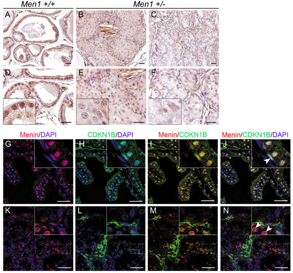
Reduced CDKN1B expression in prostate cancers from Men1+/- mice. (A-F) IHC was performed on paraffin-embedded sections of prostate tissues from Men1+/+ (A, D, n = 4) and Men1+/- (B, E and C, F, n = 4) mice using an antibody against CDKN1B. Note that in the normal prostate from aged Men1+/+ mice (26-month-old), CDKN1B is expressed in all prostatic epithelial cells, but not in all stromal cells (A, D). CDKN1B expression is reduced in an in situ carcinoma (B, E, 26-month-old mouse) and an adenocarcinoma (C, F, 23-month-old mouse) from Men1+/- mice when compared with the wild-type prostate. Panels D-F are two-fold magnifications of the upper panels (A-C). Insets show an amplified view of a part of the prostate glands. (G-N) Menin (red) and CDKN1B (green) expressions examined by double IF staining in prostate glands from a 21-month-old Men1 wild-type mouse (G-J) and an adenocarcinoma from a 23-month-old Men1+/- mouse (K-N). DAPI stains cell nuclei. Note that menin and CDKN1B expressions are co-localised in normal prostate epithelium, whereas both disappear in the cancerous lesions. Arrowheads show that basal cells remain both menin and CDKN1B positive in cancerous lesions. Boxed areas are magnified in the insets. Scale bars, 50 μm.

