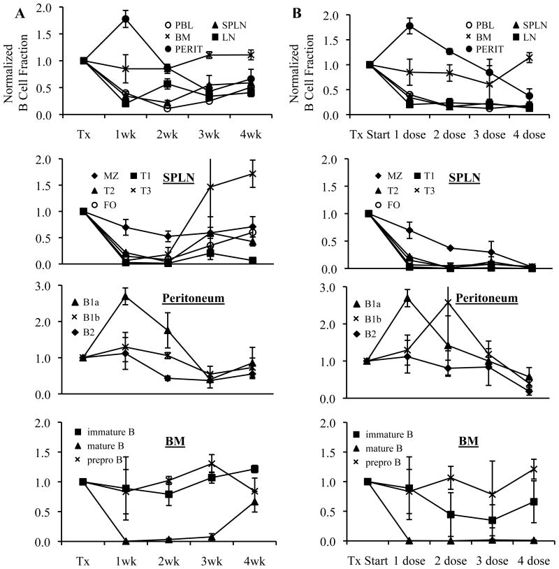
Figure 1. B cell depletion in lupus prone mice improves with prolonged treatment.
10 wk old NZB/NZWF1 mice were treated with (A) a single dose of anti-mCD20 (300 μg) or control anti-human CD20 mAb intravenously and tissues were harvested at 1,2,3,4 weeks following treatment or (B) 1,2,3 or 4 weekly doses of anti-mCD20 and tissues were harvested one week after the last antibody injection. B cells were enumerated by flow cytometry, with subset definitions as defined in the Materials and Methods. B cell subsets are depicted as a normalized ratio (mean +/− SE) compared to control treated group with n=4 animals per group and time point. PBL, peripheral blood. SPLN, spleen. LN, lymph node. BM, bone marrow. PERIT, peritoneum. MZ, marginal zone. FO, follicular.

