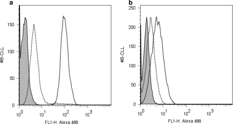
Fig. 2.
Peptide inhibition of CLL cell staining. Fluorescently labeled alemtuzumab (a) or rituximab (b) was incubated with primary CLL cells and evaluated by flow cytometry (solid line). As expected, robust staining with alemtuzumab was seen, while the staining for CD20 with rituximab was weak. When peptides pCp-1B or pRTX-10B were added at a 25,000 M excess (dashed lines), the cell labeling as largely abrogated. The shaded histogram represents CLL cells incubated with fluorescently labeled normal human IgG

