Figure 1.
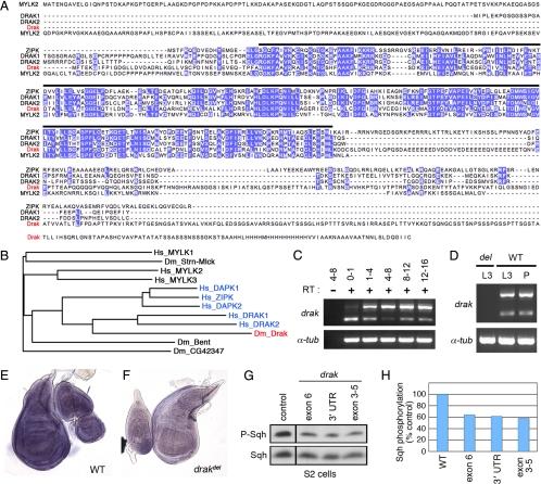
Drak is the single Drosophila homologue of DAPK family kinases. (A) Multiple sequence alignment of Drosophila melanogaster Drak with human DRAK1 and DRAK2, ZIPK, and skeletal muscle MLCK (MYLK2). Purple, identical amino acids conserved in at least three of the five sequences. Black bar, limits of the conserved kinase domains. (B) Dendrogram showing relation of Drak and other Drosophila melanogaster MLCK-like proteins to human DAPK (blue) and MLCK family kinases, based on alignment of kinase domain sequences. Hs_MYLK1, 2, and 3 correspond to nonmuscle, skeletal muscle, and cardiac Myosin light chain kinases. (C and D) Developmental time course analysis of drak expression by RT-PCR. RNA was from staged embryos (C) and third instar larval (L3) and pupal (P) wing discs (D). Numbers in C represent ages of the embryos in hours after egg laying. drak primers amplified two bands representing unspliced (upper) and spliced mRNA. Reverse transcriptase was left out of one reaction to confirm that both products derived from RNA and not contaminating genomic DNA. α-tubulin84B served as a positive control. (E and F) In situ hybridization analysis of drak expression in wing/leg/haltere imaginal discs from wild-type (E) and drakdel mutant (F) animals. (G and H) Western blot analysis of lysates from S2 cells treated with dsRNAs targeting GFP (control) or three nonoverlapping regions of the drak transcript (exons 3–5, exon 6, and 3′ untranslated region), using anti-phospho-MRLC and anti-Sqh antibodies. Levels of phospho-Sqh, normalized to total Sqh levels, are plotted in H. Phospho-Sqh images are all from the same exposure of a single blot with intervening lanes removed, as are total Sqh images.

