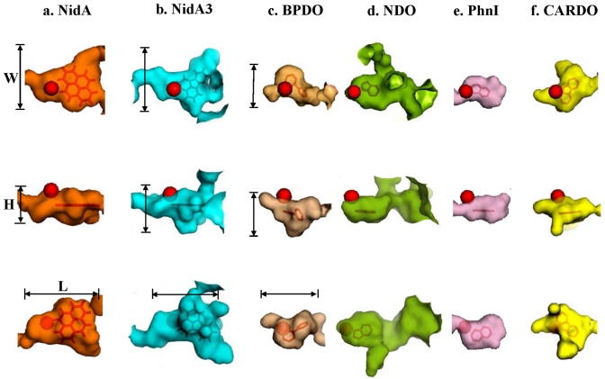
FIG 5.
Surface plots of the substrate-binding pockets of RHO enzymes bound to their substrate. The mononuclear iron is represented as a red ball. The width (W), height (H), and length (L) are shown for NidA, NidA3, and BPDO. See Table 3 for detailed numerical data for the active sites compared and their sources (microorganisms).

